
It is an intriguing finding, particularly for those interested in static magnetic field therapy. But it also raises important questions about the quality of the evidence, how different magnetic technologies compare, and what conclusions can reasonably be drawn.
Research focus: The Costantino study examined quadrupolar permanent magnets during wrist and hand fracture care. Its observations merit further investigation, but the study was preliminary and did not establish faster bone healing with Q Magnets. The article examines the evidence, its limitations, and related customer experiences.
How Do Broken Bones Normally Heal?
Bone is living tissue that continually responds to mechanical loading, biological signals and its surrounding environment.
When a fracture occurs, the body initiates a coordinated repair process involving inflammatory cells, blood vessels, cartilage-forming cells and bone-forming cells.
Although the stages overlap, fracture healing generally progresses through four phases.
Inflammation and early repair
Following a fracture, damaged blood vessels produce a local haematoma. Inflammatory cells help remove damaged tissue and release signals involved in initiating repair.
This early response is a necessary part of healing, rather than simply something to suppress.
Soft callus formation
Cells around the fracture begin producing fibrous tissue and cartilage, creating an initial bridge between the injured bone ends.
This soft callus provides early stability but does not yet have the mechanical strength of mature bone.
Hard callus formation
Osteoblasts, the cells responsible for forming new bone, contribute to replacing the soft callus with mineralised bone.
The developing hard callus progressively strengthens the fracture site.
Bone remodelling
Over subsequent months, and sometimes years, the body reorganises the newly formed bone.
Osteoclasts remove bone while osteoblasts form new tissue, gradually restoring bone architecture in response to mechanical demands.
The duration of these stages varies considerably depending on the bone involved, fracture severity, blood supply, age, general health, stability and treatment.
This is why an uncomplicated finger fracture and a complex tibial fracture cannot be expected to follow identical recovery timelines.
It is also why researchers distinguish between early callus formation, radiographic union, clinical healing and the eventual return of normal function.
What Does Research Say About Static Magnets and Fracture Healing?
One of the more directly relevant clinical investigations was published in 2007 by Costantino and colleagues at Parma Hospital in Italy.
The paper, Treatment of Wrist and Hand Fractures with Natural Magnets: Preliminary Report, examined the use of permanent quadrupolar neodymium magnets during conventional fracture immobilisation.
Unlike electrical bone stimulators, these magnets did not require a power supply or generate programmed electromagnetic pulses.
They produced a persistent static magnetic field.
The Costantino Study: Quadrupolar Magnets Inside Plaster Casts
Between 2004 and 2006, the researchers treated patients with fractures of the wrist and hand.
The study reported 40 patients, although an inconsistency in the published demographic breakdown lists four females and 37 males, totalling 41. The fracture categories themselves total 40.
The study reported 40 patients and fractures included:
- Eight distal radius fractures.
- Eighteen scaphoid fractures.
- Ten metacarpal fractures.
- Four proximal finger phalanx fractures.
The researchers used neodymium-iron-boron magnets approximately 20 mm in diameter and 5 mm thick.
Each magnet consisted of four magnetic blocks arranged with alternating north and south poles, producing a symmetrical quadrupolar static magnetic field.

The paper describes field-strength values of approximately 12,000—12,500 gauss, although the measurement location and exposure experienced by the fracture tissues are not sufficiently characterised to establish a reproducible biological dose.
The magnets were positioned within the immobilisation devices, generally near the fracture site. Conventional fracture management, including reduction and immobilisation where required, continued.
What Did the Researchers Find?
All fractures in the reported series achieved satisfactory consolidation without complications reported by the authors.
The researchers compared immobilisation periods in their patients with conventional treatment periods described in orthopaedic literature.
Some of the reported comparisons were:
| Fracture type | Average immobilisation with magnets | Historical reference | Reported difference |
|---|---|---|---|
| Distal radius, displaced without fragmentation | 21.5 days | 30 days | 28.3% shorter |
| Distal radius, displaced with fragmentation | 28 days | 37.5 days | 25.3% shorter |
| Scaphoid | 23.7 days | 45 days | 47.3% shorter |
| Distal radius, without displacement | 23 days | 30 days | 23.3% shorter |
| Metacarpal, without displacement | 20 days | 30 days | 33.3% shorter |
| Metacarpal, displaced | 22 days | 30 days | 26.6% shorter |
| Proximal phalanx, displaced | 24.6 days | 30 days | 18% shorter |
Source: Costantino et al., 2007, Table 1. Historical reference periods were not outcomes measured in a concurrent control group.

These observations are noteworthy because the researchers used permanent quadrupolar magnets rather than PEMF equipment.
However, the interpretation requires care.
What Are the Limitations of the Costantino Study?
The study is best understood as a preliminary clinical case series with historical comparisons, not a definitive controlled trial.
Several limitations affect the strength of its conclusions.
These limitations mean that the observed differences cannot confidently be attributed to the magnets.
The study nevertheless provides a useful starting point for further investigation because it documents an actual clinical application of quadrupolar permanent magnets in fracture management.
A larger, well-controlled trial would be needed to determine whether the findings are reproducible and clinically meaningful.
How Do the Magnets Used by Costantino Compare With Modern Q Magnets?
An important aspect of the Costantino study is its use of a quadrupolar permanent neodymium magnet arrangement. This is directly relevant to Q Magnets because quadrupolar magnetic configurations are central to its origins.
The magnets used in the Costantino study are made up of four bipolar magnets arranged into a quadrupolar array. Whereas Q Magnets are from a single magnetic body, but magnetized into a quadrupolar magnet.
See video explanation below.
Are Static Magnets the Same as PEMF Bone Stimulators?
No. This is one of the most important distinctions in the fracture-healing literature.
Both technologies involve magnetic fields, but they produce different physical exposures.
| Feature | Permanent static magnets | PEMF devices |
|---|---|---|
| Field generation | Permanent magnetic material | Electrically powered coils |
| Field behaviour | Approximately constant at a fixed position | Changes with time |
| Electrical power | Not required | Required |
| Exposure characteristics | Field strength, geometry, gradients and duration | Pulse waveform, frequency, intensity, duty cycle and duration |
| Clinical evidence | Limited fracture-specific human research | Larger research literature, with results varying by indication and device |
| Regulatory status | Depends on the specific device and intended use | Certain bone-growth stimulators have authorisation for specified indications |
A stationary permanent magnet produces a static field. A PEMF device produces time-varying magnetic fields, which can induce electric fields within tissue.
These are not interchangeable exposures.
Some PEMF devices have been investigated and authorised for particular bone-healing indications, including certain cases of delayed union or nonunion. That does not mean every PEMF device has demonstrated benefit for every fracture.
More importantly, evidence supporting a particular PEMF treatment cannot automatically be transferred to permanent static magnets.
The reverse is equally true.
For readers interested in these physical differences, our educational resources on static magnetic field therapy provide further context.
How Might Static Magnetic Fields Influence the Biological Processes Involved in Bone Healing?
Bone repair depends on a complex biological environment involving osteoblasts, osteoclasts, inflammatory mediators, blood supply, mechanical stability and cellular signalling.
Researchers have investigated whether electromagnetic exposures influence processes such as osteoblast differentiation, osteoclast regulation, calcium-related signalling and the expression of proteins involved in bone formation. However, findings from laboratory experiments and PEMF research cannot automatically be transferred to quadrupolar permanent magnets.
The Aliabadi Study: Microscopic Evidence of Tissue Repair
In 2012, Aliabadi and colleagues published Evaluation of the Effect of Static Magnetic Field in Treatment of Tendon Injuries in Dog in Comparative Clinical Pathology.
They investigated experimentally induced superficial digital flexor tendon injuries in ten dogs, divided into groups of five animals. The experimental group received exposure from six 800-gauss permanent magnets positioned around the injured tendon region for 30 days.
Microscopic examination and biomechanical testing identified differences between the groups. At day 30, the authors reported the following qualitative histological observations:
| Histological feature (day 30) | Control group | Static-field group |
|---|---|---|
| Inflammatory cells | Mild | Absent |
| Angiogenesis (new blood vessels) | Mild | Extensive |
| Collagen formation | Moderate | Extensive |
Source: Aliabadi et al., Table 1. These are authors’ qualitative histological grades, not quantitative vascular measurements. The sample was small (five animals per group).
The researchers also described new capillaries and better-organised collagen fibres in the magnet-exposed tendons. These microscopic observations are different from a report of pain relief alone.
Did the Treated Tendons Become Mechanically Stronger?
The authors also reported greater tendon stiffness in the exposed group and statistically significant differences, although only five animals were in each group. The measurements support an observation of changed tendon properties in this experimental model, not a conclusion about bone healing in humans.
This was an animal study of tendon tissue, not a fracture trial. Its magnetic exposure was not characterised sufficiently to determine equivalence to Costantino’s configuration or to Q Magnets.
Read our earlier article on static magnetic fields and tendon repair.
Could Angiogenesis Be a Mechanism Linking Static Magnetic Fields and Fracture Healing?
One noteworthy finding in Aliabadi’s study was the reported increase in angiogenesis: the formation of new blood vessels from existing vascular networks. Angiogenesis is essential to normal fracture repair, because vessels deliver oxygen, nutrients and signals to the developing bone callus.
Angiogenesis and osteogenesis (new bone formation) are closely coordinated. Vascular endothelial growth factor (VEGF) is one signalling molecule involved in that process. A review on VEGF and fracture healing explains the role of vascular development in bone repair.
Aliabadi’s observations suggest that static magnetic-field exposure may influence some aspects of tissue repair under experimental conditions. They raise the question of whether changes in vascular development, inflammatory signalling or collagen organisation could help explain Costantino’s preliminary observations.
This is a research hypothesis, not a demonstrated mechanism. Aliabadi studied dog tendons, not human bone, and Costantino did not measure angiogenesis, VEGF expression or related vascular endpoints in fractures. Neither study established that Q Magnets accelerate human fracture healing.
Well-controlled future studies could assess vascular development, osteoblast activity, fracture-callus formation, structural union and bone strength using reproducibly characterised static magnetic fields.
Why Field, Dose and Placement Matter
At Q Magnets, we use the Field | Dose | Placement (FDP) framework to explain the physical variables involved in applying multipolar permanent magnets.

These variables are particularly relevant when interpreting the Costantino and Aliabadi studies, which involved different magnetic configurations, tissues and application methods.
A quadrupolar magnet positioned inside a plaster cast is not necessarily equivalent to a different magnet placed externally over another part of the body.
Similarly, two magnets with the same quoted surface gauss value may produce different field distributions at depth.
These distinctions help explain why the physical details of an intervention matter when designing and comparing research.
They do not, by themselves, demonstrate an effective magnetic dose for fracture healing.
What Can Customer Experiences Tell Us About Fracture Recovery?
Alongside published research, Q Magnets has received accounts from customers and practitioners describing their experiences during recovery from fractures.
These stories are valuable because they illustrate what people notice in everyday life: discomfort, sleep, movement, work and rehabilitation.
They also need to be interpreted correctly.
A person reporting less pain after using a magnet is describing a symptomatic experience.
That is different from demonstrating that the fracture healed faster.
A Plastic Surgeon, a Marathon and Tibial Stress Fracture
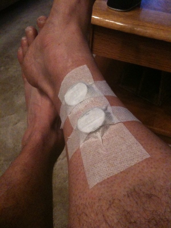
One of the earlier accounts came from Canadian plastic surgeon Dr Thibert, an enthusiastic marathon runner.
Following a tibial stress fracture during training, he became concerned that his plans to participate in an upcoming marathon might be disrupted.
After a colleague suggested magnetic therapy, he investigated Q Magnets and subsequently described a marked improvement in discomfort following their use.
He also acknowledged the importance of rest during his recovery.
He later reported returning to marathon participation.
His account is interesting both because of his medical background and because he took the time to describe his personal experience.
However, several factors need to be separated.
His reported reduction in discomfort is a personal observation. His subsequent return to running followed a period of recovery that included rest.
Neither observation establishes that Q Magnets accelerated bone union.
In particular, a rapid reduction in pain cannot demonstrate that a stress fracture has healed.

Wilma Beltrame: Comfort During Tibial Plateau Fracture Rehabilitation

Wilma Beltrame described her experience following a tibial plateau fracture, an injury involving the upper surface of the shinbone near the knee.
Her account discusses rehabilitation, pain management and the use of five Q Magnets during her recovery.
Tibial plateau fractures can require substantial rehabilitation because they involve a weight-bearing joint.
Wilma’s story illustrates the importance of comfort and symptom management during a challenging recovery period.
It does not establish whether magnetic exposure influenced the rate or quality of bone healing.
A Broken Toe: A Practitioner-Reported Experience
Practitioner Megan McMahon shared an account involving a patient recovering from a broken toe.
According to the report, the patient’s pain decreased from approximately 5/10 to 1/10 over seven days, and the patient returned to work.
This is a useful example of why pain and function are important recovery outcomes in their own right.
Nevertheless, the report does not provide objective evidence that the fracture consolidated more rapidly because of Q Magnets.
Broken Ribs: Experiences With Comfort, Sleep and Returning to Work
Two other accounts concern rib fractures.
Sharlene reported sustaining four broken ribs after a serious fall, with fractures confirmed by X-ray. Her account describes using Q Magnets for comfort and returning to work within a week.
George M. Graham also reported X-ray-confirmed rib fractures. He described a reduction in pain from 10/10 to 3/10, improved sleep and a return to work.
Rib injuries can be particularly uncomfortable because ordinary breathing, coughing and movement involve the chest wall.
These experiences therefore highlight the importance of managing discomfort during recovery.
However, neither account establishes accelerated bone repair.
Read Sharlene’s broken ribs experience
Read George Graham’s rib injury experience
These are individual customer or practitioner-reported experiences, not controlled clinical evidence. Results vary, and the reports do not demonstrate that Q Magnets treat fractures or accelerate bone healing.
Why Pain Relief Does Not Necessarily Mean a Fracture Has Healed
One of the most important lessons from fracture recovery is that pain and tissue repair do not always progress at the same rate.
Pain is influenced by many factors, including tissue injury, inflammation, mechanical loading, peripheral nerve activity and how the nervous system processes incoming signals.
Bone healing, meanwhile, involves structural repair and the gradual restoration of mechanical strength.
A person may feel substantially more comfortable while the fracture remains vulnerable to further injury.
Conversely, discomfort may persist even when satisfactory structural healing has occurred.
This distinction is particularly important with stress fractures, where returning to running or other repetitive loading too early may worsen the injury.
The practical implication is straightforward:
A reduction in pain should never be used as the sole indicator that a fracture has healed or that normal activity can safely resume.
Decisions about immobilisation, weight-bearing, rehabilitation and return to sport should be guided by an appropriately qualified healthcare professional.
For further information about the nervous system’s contribution to pain, see our article on central sensitisation and persistent pain.
Can Q Magnets Be Used During Fracture Recovery?
Q Magnets are multipolar permanent magnetic devices designed to produce localised static magnetic fields.
Their current stated intended use is to potentially provide comfort or temporary, localised relief of minor aches and pains at superficial body sites.
Q Magnets are not indicated for accelerating fracture healing, treating nonunion or replacing established fracture care.
People recovering from fractures should follow the medical advice provided for their particular injury.
This may include immobilisation, surgery, protected weight-bearing, rehabilitation and follow-up imaging.
Anyone considering using Q Magnets during fracture recovery should discuss their circumstances with their treating healthcare professional, particularly when a cast, surgical fixation or other medical device is involved.
Q Magnets should not be inserted into a cast or used in a way that interferes with prescribed treatment.
They must also be kept away from magnetically sensitive medical implants and equipment, as described in the Q Magnets instructions and application guidance.
Frequently Asked Questions
Do magnets make broken bones heal faster?
There is insufficient clinical evidence to conclude that ordinary permanent magnets or Q Magnets reliably accelerate fracture healing. The Costantino study reported promising preliminary observations with quadrupolar permanent magnets, but its uncontrolled design prevents a firm causal conclusion.
What is the difference between magnetic therapy and PEMF for fractures?
Permanent magnets produce static magnetic fields, while PEMF devices generate time-varying electromagnetic fields. Their exposure characteristics, mechanisms under investigation, clinical evidence and regulatory indications differ.
Can quadrupolar magnets stimulate bone growth?
The Costantino study investigated quadrupolar permanent magnets in fracture management, but it did not conclusively demonstrate that these magnets stimulate bone growth. Additional controlled human studies would be required.
Could angiogenesis explain how magnetic fields influence bone repair?
Possibly. Angiogenesis is essential to normal fracture healing, and Aliabadi and colleagues reported greater new blood-vessel formation in magnet-exposed canine tendons. Whether static magnetic fields influence angiogenesis in human fractures, and whether this improves bone healing, remains to be established.
Does reduced fracture pain mean the bone is healing faster?
No. Pain reduction and structural bone repair are different outcomes. A fracture may remain vulnerable even when discomfort has substantially improved.
Can I use Q Magnets instead of a cast or other fracture treatment?
No. Q Magnets are not a substitute for immobilisation, surgery, rehabilitation or other prescribed fracture care. Suspected fractures should be medically assessed.
Is the Costantino fracture study directly relevant to Q Magnets?
Yes, in terms of the underlying magnetic technology. The researchers used quadrupolar permanent neodymium magnets, belonging to the same broad technology category as quadrupolar Q Magnets. Modern Q Magnets use a refined single-body construction, and company testing has reported stronger localised gradients than the original Magna Bloc design. However, the clinical findings reported by Costantino have not been directly tested or replicated using Q Magnets.
What Can We Conclude About Magnets and Fracture Healing?
The research provides several observations that deserve further investigation.
Costantino and colleagues reported shorter immobilisation periods in patients treated with quadrupolar permanent magnets alongside conventional fracture management. This involved the same broad category of static magnetic-field technology used in quadrupolar Q Magnets, whose refined design has shown stronger localised field gradients in the company’s comparison with the original Magna Bloc arrangement.
The Aliabadi animal study adds another dimension. The authors reported increased angiogenesis, reduced inflammatory-cell presence and improved collagen organisation, alongside greater tendon strength in biomechanical testing.
These findings are interesting because vascular development, inflammatory signalling and extracellular matrix formation also contribute to normal fracture repair. Together, the studies offer a scientifically plausible reason to investigate whether carefully characterised static magnetic fields may influence bone-repair processes.
The evidence remains preliminary. Costantino lacked a concurrent control group, Aliabadi studied animal tendons, and neither study established that Q Magnets accelerate fracture healing in humans.
Q Magnets customer stories provide a perspective on comfort, sleep, pain and function during fracture recovery; they remain individual experiences rather than evidence of faster bone repair.
Controlled studies measuring fracture-callus formation, vascular changes, structural union and mechanical strength are needed. Until then, Q Magnets’ stated intended use remains temporary, localised relief of minor aches and pains.
The continuing scientific question is whether engineered static magnetic fields influence biological tissue-repair processes—and under what measurable conditions.
References and Further Reading
- Costantino C, Pogliacomi F, Passera F, Concari G. Treatment of wrist and hand fractures with natural magnets: preliminary report. Acta Biomedica. 2007;78:198—203. PubMed.
- The Effects and Mechanisms of Electromagnetic Fields on Bone Remodeling: From Clinical to Laboratory. Journal of Orthopaedic Translation, 2025. Publisher article. This review addresses electromagnetic field research more broadly and should not be interpreted as Q Magnets-specific clinical evidence.
- Q Magnets — How to Use Q Magnets.
- Q Magnets — Central Sensitisation.
- Aliabadi A, Dehghani SN, Farahmand M, Varzandian S, Dehghan A. Evaluation of the effect of static magnetic field in treatment of tendon injuries in dog. Comparative Clinical Pathology. 2013;22:1057–1060. Published online 2012. DOI: 10.1007/s00580-012-1525-3.
- Beamer B, Hettrich C, Lane J. Vascular endothelial growth factor: an essential component of angiogenesis and fracture healing. HSS J. 2010;6:85–94. PubMed.
- Q Magnets — Magna Bloc Design Comparison (device history, engineering comparison and video; company-reported findings).
- Q Magnets — Static Magnetic Fields and Tendon Repair.
Explore the Science of Static Magnetic Fields
Interested in understanding how multipolar permanent magnets differ from conventional magnetic products, and why field geometry, exposure and placement matter?
Explore our Q Magnets educational resources and learn more about the principles behind static magnetic field therapy.
For more information: Read our complete guide to magnetic therapy for pain conditions
Medical information notice: This article is provided for education and research discussion. It does not recommend Q Magnets as a treatment for fractures or impaired bone healing. Always seek appropriate medical care for a suspected or confirmed fracture.