
Central Sensitization
Central Sensitization is an increase in the excitability of neurons within the central nervous system starting at the spinal cord level. Normal inputs from everyday experiences such as touch are amplified to produce abnormally painful responses. Central Sensitization is a major cause of pain hypersensitivity.

Central Sensitization: When the Nervous System Amplifies Pain
Central sensitization is a condition in which the central nervous system becomes hypersensitive to sensory input, causing normal signals from the body to be interpreted as painful.
Instead of pain reflecting the severity of an injury, the nervous system itself becomes over-responsive, amplifying signals from the body.
This phenomenon is now recognised as one of the key biological mechanisms behind many forms of chronic pain.
In central sensitization:
• the spinal cord and brain become more excitable
• pain signals are amplified
• normal sensations such as touch can become painful
• pain may spread beyond the original injury
Central sensitization helps explain why pain can persist long after tissues have healed.
Central Sensitization and Chronic Pain
Central sensitization plays an important role in the pathophysiology of many chronic pain conditions, including:
• chronic low back pain
• fibromyalgia
• migraine
• post-herpetic neuralgia (shingles)
• diabetic neuropathy
• temporomandibular joint disorder (TMJ)
• complex regional pain syndrome (CRPS)
In these conditions, pain is no longer driven primarily by tissue damage but by changes in how the nervous system processes sensory signals.
Researchers now recognise that chronic pain often develops through a progressive cascade of nervous system changes, beginning with local tissue injury and potentially evolving into central nervous system hypersensitivity.
To understand this broader process, see our article:
Why Pain Becomes Chronic And Why Early Treatment Matters
Central Sensitization can also be a consequence of dysfunctional pain processing such as fibromyalgia, where somehow normal neural inputs from mechanotransducers get diverted into the pain pathway.
The underlying mechanisms of Central Sensitization are complex and not fully understood. They are thought to involve neurotransmitters such as glutamate and substance P that cause nerve fibres to become more “leaky” to Na+ and Ca2+ ions and hence lower the neural activation threshold.
Peripheral Sensitization and Ion Channel Proliferation
Modern pain neuroscience has also identified important changes that occur before central sensitization develops, particularly at the level of the peripheral nerves.
Research discussed in the Explain Pain framework developed by David Butler and the Neuro Orthopaedic Institute (NOI Group) describes how injured nerves undergo adaptive changes that increase their sensitivity.
One of the most important of these changes is ion channel proliferation.
Following tissue injury or nerve irritation, the body may increase the number of ion channels — particularly sodium channels — located on the cell membrane of sensory neurons.
This process may begin within days of injury and can continue for several weeks.
These additional ion channels create areas of increased electrical sensitivity along the nerve fibre.
Researchers sometimes describe these areas as abnormal impulse generating sites (AIGS).
Because ion channels control the movement of electrically charged ions across the nerve membrane, increasing their density makes the nerve easier to activate.
In practical terms, this means:
• the threshold for firing a pain signal becomes lower
• neurons become more excitable
• spontaneous nerve firing may occur
This process is commonly referred to as peripheral sensitization.
The Sensitization Cascade
Peripheral and central sensitization are best understood as parts of a connected biological process rather than completely separate conditions.
Following injury, inflammation or persistent nerve irritation, sensory neurons can become more excitable. Changes may include altered ion-channel expression and membrane behaviour, including changes involving sodium and calcium channels.
As these neurons become easier to activate, they may produce stronger, more frequent or sometimes spontaneous signals.

Within the dorsal horn of the spinal cord, repeated nociceptive input can contribute to changes in neurotransmitter activity, receptor behaviour and intracellular signalling. These changes may lower neuronal firing thresholds and amplify subsequent sensory input.
As sensitization develops, normally harmless stimuli may become painful, a phenomenon known as allodynia.
Painful stimuli may produce an exaggerated response, known as hyperalgesia.
Sensitivity may also spread beyond the original site of injury, producing secondary hyperalgesia.
These changes are examples of neuroplasticity: the nervous system adapting in response to repeated activity.
What Happens at the Cell Membrane
An important feature of sensitization is that repeated nociceptive activity is not biologically neutral.
Persistent signalling can contribute to activity-dependent changes in peripheral sensory neurons and, over time, within spinal and higher pain-processing pathways.
This provides a biological rationale for reducing excessive nociceptive input as early as practical rather than simply waiting for persistent pain pathways to become established.
That does not mean that every episode of pain will progress to central sensitization, or that reducing pain early necessarily prevents chronic pain. Many factors influence pain chronification.
However, where persistent peripheral sensitization is contributing sustained input into the nervous system, reducing that input may plausibly reduce one of the drivers involved in the sensitization process.
This is one reason early and appropriate pain management, rehabilitation and restoration of normal movement can be important.
At the microscopic level, this process can be summarized as:

Recognising Central Sensitization in Clinical Practice
Clinicians often recognise central sensitization through several key features.
Hyperalgesia
Painful stimuli produce greater than normal pain responses.
For example, a mild injury may cause disproportionate pain.
Allodynia
Normally non-painful sensations become painful.
Examples include:
• light touch
• clothing brushing the skin
• gentle pressure
Secondary Hyperalgesia
Pain sensitivity spreads to areas surrounding the original injury, even where tissue damage has not occurred.
Persistent Pain
Pain continues long after tissue healing should have occurred.
(Nijs, Van Houdenhove et al. 2010) provided guidelines for manual therapists to recognise central sensitization in patients with musculoskeletal pain. The examination included for instance…
1. Assessment of pressure pain thresholds at sites remote from the symptomatic site;
2. Assessment of sensitivity to touch during manual palpation at sites remote from the symptomatic site; and
3. Assessment of pressure pain thresholds during and following exercise
(Nijs, Meeus et al. 2011) provided a list of treatment options for the treatment of central sensitization, which included pharmacotherapy such as acetaminophen (paracetamol), serotonin-reuptake inhibitor drugs, opioids, NMDA receptor blockers and Ca2+ channel ligands such as pregabalin and gabapentin.
“Just like context-dependant drugs…”
Laboratory studies and clinical observations have raised the possibility that appropriately configured static magnetic fields may have greater relevance where sensory neurons are sensitized or firing persistently.
Research using cultured sensory neurons found that steep static magnetic field gradients could reversibly alter neuronal excitability and suppress sustained action-potential firing under experimental conditions.
This provides a plausible mechanistic basis for investigating whether localized static magnetic field gradients may influence sensitized sensory-nerve activity.
Magnetic Field Gradients and Nerve Excitability
Research over several decades has investigated whether particular static magnetic field configurations can influence sensory-neuron activity.
Laboratory studies conducted by researchers associated with Vanderbilt University examined steep magnetic field gradients generated by multipolar magnetic arrays.
In cultured sensory neurons, exposure to particular static magnetic field gradients was associated with reversible changes in neuronal excitability and action-potential firing.
In one experiment, sensory neurons exposed to capsaicin developed sustained firing. Exposure to the magnetic field gradient temporarily suppressed this firing, with activity returning after the field was removed.
The findings support a possible reversible neuromodulation mechanism rather than permanent nerve suppression.
One proposed explanation involves changes in membrane behaviour and sodium and calcium ion permeability, although the precise biological mechanism remains unresolved.
The clinical significance of these laboratory findings requires further investigation.
Magnetic Therapy and Targeted Neuromodulation
Unlike systemic pharmacological treatments, magnetic therapy devices are typically applied locally over specific anatomical targets.
One advantage of wearable magnetic devices is that they can provide continuous exposure to the therapeutic field without interfering with normal daily activities.
In clinical practice, magnetic devices are sometimes placed:
• directly over painful tissues
• over spinal segments associated with the affected nerves
• over trigger points or sensitized regions
Correct device design and placement are critical, as the therapeutic effect depends on the characteristics of the magnetic field gradient reaching the target tissue.
Clinical Observations
Clinicians working with patients experiencing chronic pain have reported that when magnetic field therapy is correctly applied, some patients experience noticeable reductions in pain sensitivity.
In certain cases, improvements have included:
• decreased hypersensitivity to touch
• improved tolerance to movement or manual therapy
• reductions in background pain levels
While these observations require further scientific investigation, the low risk associated with non-invasive magnetic therapy makes it an area of ongoing clinical interest.
After 10 years of research including in-vitro cell studies and randomised controlled trials the group of neurologists at Vanderbilt Medical University lead by Dr Robert Holcomb, M.D., Ph.D. concluded that the evidence suggested that the most likely mechanism of action is that the steep field gradients generated by the quadrapolar magnetic field is altering nerve excitability as a result of changes in membrane permeability to Na+ and Ca2+ ions (McLean, Holcomb et al. 1991), (McLean, Holcomb et al. 1995), (Cavopol, Wamil et al. 1995), (McLean, Engstrom et al. 2001).
(McLean, Engstrom et al. 2001) describes the cell study where nerve fibres were exposed to the noxious stimuli capsaicin and after 5 minutes of exposure to the gradient of the Quadrapolar array, the action potential firing was totally blocked and fully recovered 10 minutes after the removal of the field.
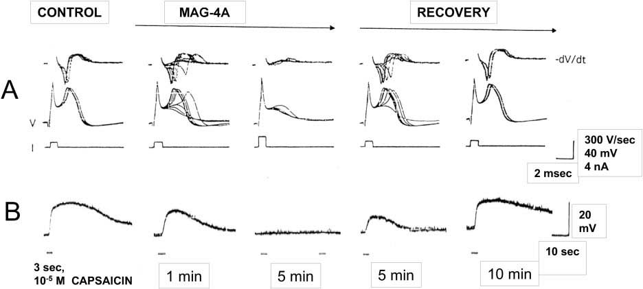
In a double blind, randomised controlled trial (Vallbona, Hazlewood et al. 1997) assessed the effectiveness of a concentric alternating multipolar rubberised magnet which produces a magnetic field gradient, albeit most likely with a penetration of less than 5 mm. Of the 50 post-polio patients enrolled in the study, 29 were in the active group and 21 the inactive. The devices were placed over the most painful trigger point for 45 minutes. Those who received the active device reported significantly less pain than those that received the sham device.
As an indication of the therapeutic effects of an inhomogeneous static magnetic field on peripheral sensitization, Kovacs-Balint et al (2011) conducted a double-blind, placebo controlled trial and demonstrated that exposure to an optimized field significantly increased the thermal pain threshold in healthy young adults.
What is clear when it comes to magnetic therapy is that unless you are using a magnetic device with an optimised design, with the right field characteristics, correctly placed so that the penetrating field envelopes the offending target tissue, then the therapy is unlikely to be effective.
With the recent release of the third generation Q Magnet models it is now possible to apply a device with an optimised therapeutic field that will envelope the target tissue. Specifically when it comes to central sensitization, the most important devices for the lumbar spine are QF28-6 and QF28-3 and where there are two adjacent levels indicated, usually L4/5 and L5/S1 the OF50-3. The OF50-3 model is a powerful rare earth neodymium octapolar magnet with a 50mm diameter that can envelope adjacent levels of the spinal cord with a magnetic field gradient, which to our knowledge is the first time this has been achieved with a static magnet device.
What is encouraging for practitioners working with complex chronic pain conditions is that clinical observations suggest that when Q magnet therapy is correctly applied, reductions in hypersensitivity and improvements in symptoms may sometimes occur quite rapidly.
While the scientific evidence for these observations requires much more research, the advantage for the patient is that the risk of intervention and expense is almost negligible.
Understanding the Bigger Picture of Chronic Pain
Central sensitization is only one stage within a broader process through which pain can become chronic.
Researchers now describe a pain chronification cascade in which:
- Tissue injury triggers peripheral sensitization, partly driven by ion channel proliferation on nerve membranes.
- Persistent peripheral signals may then lead to central sensitization within the spinal cord.
- Over time, brain networks involved in pain processing can also reorganize, further reinforcing chronic pain.
Emerging research is examining whether persistent pain involves broader changes in the organisation of neural activity across the brain.
For example, Khatib, Robinson and Yassin (2026) have proposed that chronic primary pain may involve disrupted thalamocortical oscillatory activity and abnormal organisation of endogenous electromagnetic neural activity.
Their proposal remains a hypothesis, and the authors acknowledge that its proposed causal hierarchy has not yet been demonstrated.
However, it reflects a broader shift in pain science: persistent pain is increasingly understood not simply as a signal coming from damaged tissue, but as a condition involving changes in neuronal excitability, connectivity and nervous-system processing.
This reinforces an important principle:
The longer abnormal pain signalling persists, the more the nervous system itself may become part of the problem.
Understanding these stages helps explain why early treatment of pain and injury is important.
To learn more about this broader framework, read:
Why Pain Becomes Chronic And Why Early Treatment Matters

Case Studies that demonstrate the effects of Q magnet therapy on Central Sensitization.
CASE STUDY 1 – By Lifestyle Therapies in Brisbane, Australia
Sam, a fifty year old man with a 15 year history of chronic lower back pain was referred to Lifestyle Therapies from a local health provider. Sam had been suffering with constant deep pain and numbness in his right foot for 15 years, initially injuring his back when he was 20 years old. In his thirties, Sam underwent a laminectomy, followed by lumbar fusion of L5-S1 a few years later. Since then he has not lived a day without pain, significantly affecting his quality of life. In addition Sam was suffering hyperalgesia in his lower back due to an incident with a TENS machine. His skin in the lumbar spine was so sensitive, wearing clothes was unbearable which was an indication of Central Sensitization. He was unable to sit in a car for more than one hour without suffering significantly. Sam had one goal, which was to return to his hobby of painting.
- Initial assessment findings:
- Muscle inhibition of core muscles
- High tones in gluteal muscles
- Tender to touch adductors, ITB, and Quadricep muscles.
The lumbar region was hypersensitive to pain, even with light touch; a sign of static mechanical allodynia.
Sam was fitted with the OF50-3 Octapolar Q Magnet. This neuromagnetic device was selected since it’s large enough to cover the adjoining lumbar vertebrae L4/5 and L5/S1.
One week after seeing Sam, he reported having pain-free moments for the first time in 14 years. He was also able to tolerate the drive from his house to the clinic with minimal pain. During the next week Sam was disappointed that his symptoms were returning. After examining the Q Magnet, he realised it had been reattached incorrectly, with the sticker and flux plate facing the skin. Once corrected, his symptoms immediately began to subside.
After seven sessions, the practitioner reported that Sam’s hypersensitivity had reduced substantially and that he was able to sit and paint for five hours. Light touch was reportedly no longer painful.
This reduction in allodynia was consistent with reduced pain sensitivity, although the observation alone cannot establish whether the change occurred predominantly at peripheral, spinal or higher levels of the nervous system.
CASE STUDY 2 – By Edwards Physiotherapy in Auckland, New Zealand
JC is a medical doctor and had sufferred with chronic backache for about 14 years with varying degrees of pain and stiffness, with pins and needles. When the pain was particularly bad, JC would feel lethargic and down, and have difficulty sleeping and have to take pain-killers to find some relief. JC tried all sorts of treatment: physiotherapy, chiropractic, osteopathy, neurolink, Chinese and Western acupuncture, breathing exercises, etc. Most forms of treatment would only give me temporary relief.
JC was referred to Edwards Physiotherapy and was introduced to Q Magnets. JC felt much less pain in just 15 minutes after the QF28-3 was applied and improved again when the OF50-3 was applied. For the first time in over a decade, JC can go through her day feeling quite comfortable while wearing the OF50-3. JC now responds well to the Feldenkrais treatment, which she had been seeking in the previous two years and is hopeful that one day she will be completely healed.
REFERENCES:
Butler, D., Moseley, L. (2003). Explain Pain. Adelaide, Australia: Neuro Orthopaedic Institute (NOI Group).
Cavopol, A. V., A. W. Wamil, et al. (1995). “Measurement and analysis of static magnetic fields that block action potentials in cultured neurons.” Bioelectromagnetics 16(3): 197-206.
Kovacs-Balint, Z., A. Csatho, et al. (2011). “Exposure to an inhomogeneous static magnetic field increases thermal pain threshold in healthy human volunteers.” Bioelectromagnetics 32(2): 131-139.
Lyndon, S. (2026). Pathophysiology of Chronic Pain. Clin J Pain. doi:10.1097/AJP.0000000000001362. PMID: 41614224.
McLean, M., S. Engstrom, et al. (2001). “Static Magnetic Fields for the Treatment of Pain.” Epilepsy & Behavior 2(3): S74-S80
McLean, M. J., R. R. Holcomb, et al. (1991). “Effects of Steady Magnetic Fields on Action Potentials of Sensory Neurons in Vitro.” Environmentalist 8(2).
McLean, M. J., R. R. Holcomb, et al. (1995). “Blockade of sensory neuron action potentials by a static magnetic field in the 10 mT range.” Bioelectromagnetics 16(1): 20-32.
Nijs, J., M. Meeus, et al. (2011). “Treatment of central sensitization in patients with ‘unexplained’ chronic pain: what options do we have?” Expert Opin Pharmacother 12(7): 1087-1098.
Nijs, J., B. Van Houdenhove, et al. (2010). “Recognition of central sensitization in patients with musculoskeletal pain: Application of pain neurophysiology in manual therapy practice.” Man Ther 15(2): 135-141.
Vallbona, C., C. F. Hazlewood, et al. (1997). “Response of pain to static magnetic fields in postpolio patients: a double-blind pilot study.” Arch Phys Med Rehabil 78(11): 1200-1203.
Khatib, M., D. Robinson, et al. (2026). “A bioelectromagnetic hypothesis of chronic primary pain: from thalamocortical dysrhythmia to the consciousness-brain interface.” Frontiers in Pain Research 7: 1790293. doi:10.3389/fpain.2026.1790293.
FAQs
Frequently Asked Questions About Central Sensitization
What is central sensitization?
Central sensitization is a condition in which the central nervous system becomes hypersensitive to sensory signals.
Instead of pain reflecting the severity of a tissue injury, the spinal cord and brain begin amplifying incoming sensory information.
As a result, normal stimuli may be perceived as painful, and painful stimuli may feel more intense than expected.
Central sensitization is now recognised as a key mechanism involved in many forms of chronic pain.
What causes central sensitization?
Central sensitization usually develops after persistent or repeated activation of pain pathways.
Common triggers include:
• injury or tissue inflammation
• nerve irritation or damage
• ongoing musculoskeletal strain
• repeated painful stimulation
When these signals continue over time, neurons in the spinal cord and brain can become more excitable.
What is peripheral sensitization?
Peripheral sensitization refers to increased sensitivity of nerves near the site of an injury.
Following tissue damage, inflammatory chemicals and cellular changes can make sensory nerves more responsive to stimulation.
One important mechanism involves ion channel proliferation on the nerve cell membrane, particularly sodium channels.
What is hyperalgesia?
Hyperalgesia is a condition in which painful stimuli produce a stronger-than-normal pain response.
What is allodynia?
Allodynia occurs when normally non-painful sensations become painful.
Examples include:
• light touch
• clothing brushing against the body
• gentle pressure
Can central sensitization be reversed?
In many cases, central sensitization can improve over time because the nervous system is capable of neuroplastic change.
Treatment approaches may include:
• pain neuroscience education
• graded movement or exercise
• cognitive behavioural strategies
• neuromodulation therapies
• appropriate medical management
-
 Q Magnets General Use SetAU$285.00
-
 Q Magnets Complex Lower Back SetAU$525.00
-
 Q Magnets Lower Back SetAU$336.00
-
 Q Magnets Active Athlete SetAU$667.00
-
 Q Magnets QB13 Blanket - SmallAU$725.00

Got Q Magnets ?
We are dedicated to support our products with sound advice from experienced health professionals to achieve the best results!