While maybe not so relevant to how static magnetic fields can assist pain relief or injury recovery, this study by Lew (2019) nonetheless, does give an important insight into biological effects.
When tooth decay advances to the inner pulp, it can require extraction or root canal work to preserve the tooth.
Conservative alternatives include direct pulp capping or partial pulpotomy.
Either way, increasing the formation of dentine within the tooth is important, a process called dentinogenesis.
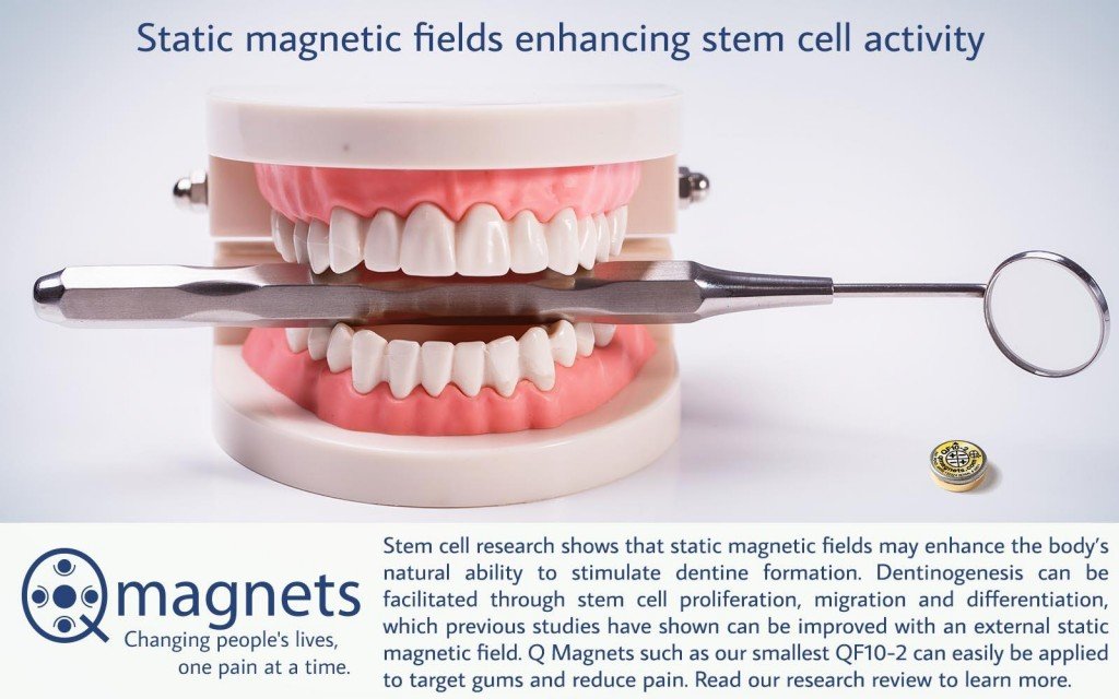
Dentinogenesis can be facilitated through stem cell proliferation, migration and differentiation, which previous studies have shown can be improved with an external static magnetic field (SMF) (Chiu 2007 & Kim 2015).
This study isolated dentine-pulp stem cells (DPSCs) from donor teeth and with the application of a 0.4 mTesla (4,000 Gauss) SMF from the north pole of a neodymium magnet, was able to assess its effects on important biological processes.
NOTE: This is approximately the same strength as the neodymium magnetic material for Q Magnets, but this is a simple bipolar magnet, not a multipolar magnet like quadrapolar or hexapolar. After simulating a pulp tissue injury, experiments were conducted to analyse the DPSC response by way of cell proliferation and migration.
Compared to the control, results of the study showed the magnetic field treated DPSCs showed statistically significant improvements in…
- Dentine-pulp stem cell migration
- Dentinogenesis (dentine formation)
Researchers suggested that the biological effects on the cells could be attributed to the static magnetic field’s effect on the molecular structure of cell membranes and Ca2+ flow. It would be reasonable to conclude that the DPSCs can sense the physical force of the SMF through the cell membrane’s biosensors. These signals are then taken from the membrane (by way of mechanotransduction) and transmitted to the cytoskeleton inside of the cell and thereby affect its functionality.
In a similar way, SMFs have been shown to significantly inhibit pro-inflammatory cytokines (interleukins, tumor necrosis factor) released from inflammatory cells. In addition, applying a moderate SMF immediately after an inflammatory injury can significantly reduce oedema (Morris 2008).
To make it practical, exposing the injured pulpal tissue to a SMF could be done by including a magnet in an occlusal splint as cleverly done by this dentist from Portugal with an QF10-2 Q Magnet. This method may enhance the body’s natural ability to stimulate dentine formation.
REFERENCES:
Chiu KH, Ou KL, Lee SY et al. (2007) Static magnetic fields promote osteoblast-like cells differentiation via increasing the membrane rigidity. Annals of Biomedical Engineering 35, 1931–9. PMID: 17721730
Kim EC, Leesungbok R, Lee SW et al. (2015) Effects of moderate intensity static magnetic fields on human bone marrow-derived mesenchymal stem cells. Bioelectromagnetics 36, 267–76. PMID: 25808160
Lew W.Z. et al (2019). Use of 0.4-Tesla static magnetic field to promote reparative dentine formation of dental pulp stem cells through activation of p38 MAPK signalling pathway. Int Endod J. 2019 Jan;52(1):28-43. PMID: 29869795, DOI
Morris et al. (2008) Acute Exposure to a Moderate Strength Static Magnetic Field Reduces Edema Formation In Rats. Am J Physiol Heart Circ Physiol: 2008 Jan;294(1):H50-7. PMID: 17982018; doi.

